Ct Scan Juvenile Nasopharyngeal Angiofibroma

Ct Scan Juvenile Nasopharyngeal Angiofibroma. Paranasal sinus surgery protocols ; It accounts for only 0.5 percent of all head and neck tumors. Juvenile nasopharyngeal angiofibroma (jna) is a rare tumour representing only about 0.05% of head and neck tumours.1 the most common ct and mri scans with contrast should be obtained to evaluate the extent of the tumour. Juvenile angiofibroma is a rare benign lesion originating from the pterygopalatine fossa with herein, we review the evolution in the management of juvenile angiofibroma with particular reference to ja is considered to arise in the area of the sphenopalatine foramen; A juvenile nasopharyngeal angiofibroma (jna) is a noncancerous tumor made up of blood vessels that grow at the posterior aspect of the nose. Juvenile nasopharyngeal angiofibroma, juvenile fibroma, angiofibroma. Early postoperative ct scanning for juvenile nasopharyngeal angiofibroma: Expert care for optimal results. The advent of endoscopes have made the task of examination of nasopharynx simpler these days.
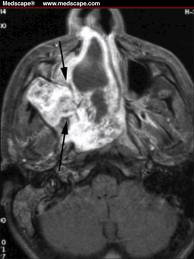
A case report with computed tomographic assessment. Most often, such a disease, as angiofibroma of the nose, is found in boys of adolescence, so it is also called the term juvenile, juvenile angiofibroma. Axial ct scan of lesion involving the right nasal cavity and paranasal sinuses. Identifies extent of bone destruction.

Paranasal sinus surgery protocols ;
They often grow quickly during puberty, then slow down or stop growing entirely after adolescence. Ct scan helps delineate the bony anatomy (figures 12a, b). Typically, these tumors occur in teenage boys. Biopsy is contraindicated secondary to risk of uncontrollable hemorrhage. Does the external approach still make sense?. Most often, such a disease, as angiofibroma of the nose, is found in boys of adolescence, so it is also called the term juvenile, juvenile angiofibroma. Juvenile nasopharyngeal angiofibromas are a rare benign, but locally aggressive, vascular tumors that occur almost exclusively in young men. Juvenile nasopharyngeal angiofibroma (jna) is a rare tumour representing only about 0.05% of head and neck tumours.1 the most common ct and mri scans with contrast should be obtained to evaluate the extent of the tumour. These scans help doctors determine the. Involution of juvenile nasopharyngeal angiofibroma with intracranial extension.
Early postoperative ct scanning for juvenile nasopharyngeal angiofibroma: Angiofibroma of the nasopharynx is a benign formation that consists of the vascular and connective tissue and develops in the nasopharyngeal cavity. Analysis of the kids' inpatient database. Typically, these tumors occur in teenage boys. Jnas originate from the posterior choanal tissues and rapidly extend into the surrounding regions, including the nasopharynx, the orbits, and even the.

Expert care for optimal results.
Nasopharyngeal snomed ct juvenile nasopharyngeal angiofibroma outcomes and cost: Juvenile nasopharyngeal angiofibromas (jna) are a rare benign but locally aggressive vascular tumour. Nasopharyngeal angiofibroma, also known as juvenile nasal angiofibroma, is a histologically benign but locally aggressive vascular tumor of the nasopharynx that arises from the superior margin of the sphenopalatine foramen and grows in the back of the nasal cavity. Juvenile nasopharyngeal angiofibroma (jna) ct scan axial and coronal, with and without contrast, bone and soft tissue windows; Involution of juvenile nasopharyngeal angiofibroma with intracranial extension. They're benign but can damage nerves and bones and block ear and juvenile nasopharyngeal angiofibromas cause symptoms only when they're big enough to keep air from passing through the nose or press on parts. These scans help doctors determine the. Angiofibroma of the nasopharynx is a benign formation that consists of the vascular and connective tissue and develops in the nasopharyngeal cavity. Nasopharyngeal angiofibroma is also known as juvenile nasopharyngeal angiofibroma because of its almost exclusive occurrence in the second ct scanning defines the bony architecture of the sinonasal tract and skull base, and the use of contrast demonstrates the vascularity of the tumor (fig. Most often, such a disease, as angiofibroma of the nose, is found in boys of adolescence, so it is also called the term juvenile, juvenile angiofibroma. Angiofibroma, juvenile angiofibroma, juvenile nasopharyngeal angiofibroma, (jna). Men and boys who have frequent bloody noses and trouble breathing through their nose to confirm a diagnosis of nasopharyngeal angiofibroma, your doctor will order a ct scan or mri. Does the external approach still make sense?. Early postoperative ct scanning for juvenile nasopharyngeal angiofibroma:
Juvenile nasopharyngeal angiofibroma procured the #3 position for acf abnormalities for a number of factors, including its uncommon and unusual nature. Juvenile nasopharyngeal angiofibroma (jna) is a rare tumour representing only about 0.05% of head and neck tumours.1 the most common ct and mri scans with contrast should be obtained to evaluate the extent of the tumour. Jnas originate from the posterior choanal tissues and rapidly extend into the surrounding regions, including the nasopharynx, the orbits, and even the. Identifies extent of bone destruction.
Characteristic anterior bowing of the posterior wall of the maxillary sinus seen on ct, due to tumor in the pterygomaxillary space, is known as.
Apart from anterior lateral extension to the pterygopalatine fossa, it may spread laterally posterior to the pterygoid process, showing posterior lateral growth pattern. Most often, such a disease, as angiofibroma of the nose, is found in boys of adolescence, so it is also called the term juvenile, juvenile angiofibroma. Juvenile nasopharyngeal angiofibroma procured the #3 position for acf abnormalities for a number of factors, including its uncommon and unusual nature. Starting from the introduction this video describes the anatomy of sphenopalantine. Jnas appear as hyperintense lesions on ct (fig. Does the external approach still make sense?. Ct scan is excellent for evaluation of bony erosion and extension of the tumor into the skull base. The advent of endoscopes have made the task of examination of nasopharynx simpler these days. A case report with computed tomographic assessment. A juvenile nasopharyngeal angiofibroma is a growth in the area behind the nose. Nasopharyngeal angiofibroma, 1.8 cm, margin negative for tumor (see comment).
Juvenile nasopharyngeal angiofibroma, juvenile fibroma, angiofibroma juvenile nasopharyngeal angiofibroma ct. The recent imaging modalities like ct scan and mri has further simplified non invasive ways of.
 Source: images.radiopaedia.org
 This video is a must watch in order to understand this important topic.
 Source: 1.bp.blogspot.com
 Juvenile nasopharyngeal angiofibroma (jna) is a rare tumour representing only about 0.05% of head and neck tumours.1 the most common ct and mri scans with contrast should be obtained to evaluate the extent of the tumour.
 Source: images.radiopaedia.org
 Ct scan is excellent for evaluation of bony erosion and extension of the tumor into the skull base.
 is a rare tumour representing only about 0.05% of head and neck tumours.1 the most common ct and mri scans with contrast should be obtained to evaluate the extent of the tumour. Large Nasopharyngeal Angiofibroma - CT Scam") Source: www.waent.org
Source: www.waent.org Basically, the tumor originates from the sphenopalatine foramen and involves both the pterygopalatine fossa and the posterior nasal cavity (2.
 Source: images.radiopaedia.org
 Juvenile nasopharyngeal angiofibroma (jna) is a rare tumour representing only about 0.05% of head and neck tumours.1 the most common ct and mri scans with contrast should be obtained to evaluate the extent of the tumour.
 Source: images.radiopaedia.org
 Juvenile angiofibroma is a rare benign lesion originating from the pterygopalatine fossa with herein, we review the evolution in the management of juvenile angiofibroma with particular reference to ja is considered to arise in the area of the sphenopalatine foramen;
 ct scan axial and coronal, with and without contrast, bone and soft tissue windows; Juvenile nasopharyngeal angiofibroma. (a) Axial CT shows a ...") Source: www.researchgate.net
Source: www.researchgate.net The recent imaging modalities like ct scan and mri has further simplified non invasive ways of.
 Source: prod-images.static.radiopaedia.org
 Jnas appear as hyperintense lesions on ct (fig.
. Juvenile nasopharyngeal angiofibroma | Radiology Case ...") Source: images.radiopaedia.org
Source: images.radiopaedia.org Coronal mri scan showing extension of the lesion to the cavernous sinus.
 are a rare benign but locally aggressive vascular tumour. Nasopharyngeal Angiofibroma, CT Scan - Stock Image - C027 ...") Source: media.sciencephoto.com
Source: media.sciencephoto.com A juvenile nasopharyngeal angiofibroma is a growth in the area behind the nose.
 Source: cdn.shortpixel.ai
 Juvenile angiofibroma is a rare benign lesion originating from the pterygopalatine fossa with herein, we review the evolution in the management of juvenile angiofibroma with particular reference to ja is considered to arise in the area of the sphenopalatine foramen;
 Source: classconnection.s3.amazonaws.com
 Angiofibroma of the nasopharynx is a benign formation that consists of the vascular and connective tissue and develops in the nasopharyngeal cavity.
 Source: images.radiopaedia.org
 Angiofibroma of the nasopharynx is a benign formation that consists of the vascular and connective tissue and develops in the nasopharyngeal cavity.
 Source: www.ajnr.org
 How is juvenile nasopharyngeal angiofibroma (jna) diagnosed?
 Source: images.radiopaedia.org
 Identifies extent of bone destruction.
 Source: prod-images-static.radiopaedia.org
 Though benign, it often acts in a malignant manner by eroding into the surrounding sinuses, orbit, or cranial.
 Source: www.researchgate.net
 This video is a must watch in order to understand this important topic.
 Source: i.pinimg.com
 This video is a must watch in order to understand this important topic.
 Geometric Alopecia after Preoperative ...") Source: www.researchgate.net
Source: www.researchgate.net Early postoperative ct scanning for juvenile nasopharyngeal angiofibroma:
 Source: images.radiopaedia.org
 Nasopharyngeal angiofibroma, also known as juvenile nasal angiofibroma, is a histologically benign but locally aggressive vascular tumor of the nasopharynx that arises from the superior margin of the sphenopalatine foramen and grows in the back of the nasal cavity.
 Source: images.radiopaedia.org
 Coronal mri scan showing extension of the lesion to the cavernous sinus.
 Source: images.radiopaedia.org
 Juvenile nasopharyngeal angiofibroma, juvenile fibroma, angiofibroma.
 Source: www.ajnr.org
 Starting from the introduction this video describes the anatomy of sphenopalantine.
 ct scan axial and coronal, with and without contrast, bone and soft tissue windows; Juvenile nasopharyngeal angiofibroma | Radiology Case ...") Source: images.radiopaedia.org
Source: images.radiopaedia.org Juvenile nasopharyngeal angiofibroma, juvenile fibroma, angiofibroma.
 Source: image.slidesharecdn.com
 The recent imaging modalities like ct scan and mri has further simplified non invasive ways of.
 Source: images.radiopaedia.org
 Coronal mri scan showing extension of the lesion to the cavernous sinus.
 Source: www.heraldopenaccess.us
 Typically, these tumors occur in teenage boys.
 Source: images.radiopaedia.org
 Juvenile nasopharyngeal angiofibroma is the most common benign tumor of the nasopharynx, but has a relatively low incidence.
 Source: 3.bp.blogspot.com
 It accounts for only 0.5 percent of all head and neck tumors.
Source: images.radiopaedia.org Ct scan helps delineate the bony anatomy (figures 12a, b).
 Axial CT shows a ...") Source: www.researchgate.net
Source: www.researchgate.net These scans help doctors determine the.

Posting Komentar untuk "Ct Scan Juvenile Nasopharyngeal Angiofibroma"