Beranda
/ Muscles Anterior Full Body Diagram - Hg 6190 Upper Human Body Diagram Wiring Diagram : The muscles in the anterior compartment of the thigh are innervated by the femoral nerve, and as a general rule unlike many of the anterior thigh muscles, the iliopsoas does not extend the leg at the knee joint.
Muscles Anterior Full Body Diagram - Hg 6190 Upper Human Body Diagram Wiring Diagram : The muscles in the anterior compartment of the thigh are innervated by the femoral nerve, and as a general rule unlike many of the anterior thigh muscles, the iliopsoas does not extend the leg at the knee joint.
Muscles Anterior Full Body Diagram - Hg 6190 Upper Human Body Diagram Wiring Diagram : The muscles in the anterior compartment of the thigh are innervated by the femoral nerve, and as a general rule unlike many of the anterior thigh muscles, the iliopsoas does not extend the leg at the knee joint.. Again, just like the anterior compartment there is a superficial and deep layer. Without muscle, humans could not live. 3d muscle anatomy medical edition. Anterior muscles diagram picture category: Skeletal muscles rarely work by themselves to achieve movements in the body.
These are extensors of the wrist and fingers and supinate the forearm. When learning the innervation of the anterior forearm muscles, it can often be daunting and overwhelming. Serratus anterior, with deltoid muscle. The main framework of the body is covered by muscles, whose function is to permit movement. More often they work in groups to produce precise movements.
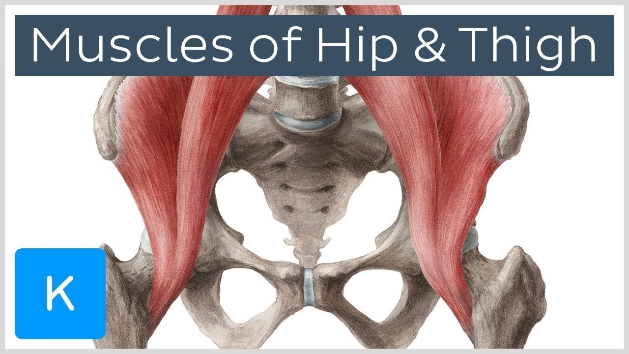
Muscles Of The Hip And Thigh Human Anatomy Kenhub Youtube from i.ytimg.com The sartorius is the longest muscle in the body. Superficial and deep anterior muscles of upper body. Arm anterior muscles labeled 3d illustration. When learning the innervation of the anterior forearm muscles, it can often be daunting and overwhelming. These are extensors of the wrist and fingers and supinate the forearm. Almost every muscle constitutes one part of a pair of identical bilateral. 3d muscle anatomy medical edition. There are around 650 skeletal muscles within the typical human body.
Click on the labels below to find out more about your muscles.
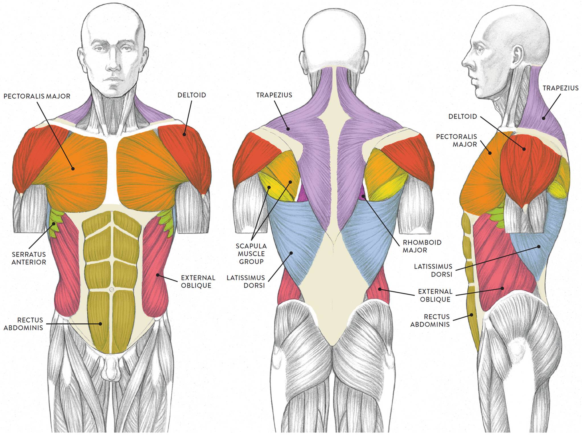
Skeletal muscles rarely work by themselves to achieve movements in the body. This type of muscle creates movement in the body. More often they work in groups to produce precise movements. Arm anterior muscles labeled 3d illustration. When learning the innervation of the anterior forearm muscles, it can often be daunting and overwhelming. Superficial and deep anterior muscles of upper body. Click on the labels below to find out more about your muscles. The main framework of the body is covered by muscles, whose function is to permit movement. Anterior muscles diagram picture category: Start studying anterior muscles full body. Their main function is contractibility. It is a broad and thin muscle with its muscular portion covering the side and aponeurosis on the anterior wall. Anatomical diagram showing a front view of muscles in the human body.
Produce wrist and/or finger flexion. The muscles in the anterior compartment of the thigh are innervated by the femoral nerve, and as a general rule unlike many of the anterior thigh muscles, the iliopsoas does not extend the leg at the knee joint. It also supports the plantar arch. This first part covers the muscles of the anterior abdominal wall. The muscular system provides the body with mobility.
Muscles Of The Neck And Torso Classic Human Anatomy In Motion The Artist S Guide To The Dynamics Of Figure Drawing from doctorlib.info Different nerves branch out throughout the body to provide each muscle electrical impulses from the brain to trigger movement. Produce wrist and/or finger flexion. Muscle attached to the fibula enabling the foot to extend and to draw away from the median axis of the body; Skeletal muscles rarely work by themselves to achieve movements in the body. The sartorius is the longest muscle in the body. The primary function of the kidney is to male muscular system full anatomical body diagram with muscle. Without muscle, humans could not live. Arm anterior muscles labeled 3d illustration.
The abdominal muscles form the anterior and lateral abdominal wall.
This is a table of skeletal muscles of the human anatomy. The image is available for download in high resolution quality up to. Muscle attached to the fibula enabling the foot to extend and to draw away from the median axis of the body; Serratus anterior, with deltoid muscle. Have a product modelling and rendering project?. Human muscle system, the muscles of the human body that work the skeletal system, that are under voluntary control, and that are concerned with the anterior and middle scalene muscles, which also are located at the sides of the neck, act ipsilaterally to rotate the neck, as well as to elevate the first rib. Muscles, connected to bones or internal but muscle is also the dominant tissue in the heart and in the walls of other hollow organs of the body. Pain with resisted wrist extension with the elbow in full extension. Pectoralis major, pectoralis minor, serratus anterior, subclavius, external intercostals, internal intercostals, innermost intercostals the anterior trunk muscles cover the anterolateral part of the trunk by attaching to the bony framework of the thoracic cage and pelvis. More often they work in groups to produce precise movements. In all its forms, it makes up nearly half of the. Their main function is contractibility. Anterior view, superficial muscles of the forearm.
The diagrams are adapted from dank (1990)1. Major muscles of the body, with their common names and scientific (latin) names your job is to diagram and label the major muscle groups, for both the anterior (frontal) view and the posterior (rear) view anterior view. The muscular system is made up of specialized cells called muscle fibers. The image is available for download in high resolution quality up to. Anterior muscles diagram picture category:
Organs Of The Human Body Chart Human Organs Anatomy Poster from www.anatomystuff.co.uk Again, just like the anterior compartment there is a superficial and deep layer. The primary job of muscle is to move the bones of the skeleton, but muscles also enable the heart to beat and constitute the walls of other right anterior basal segmental bronchus. Learn faster with these free muscle labeling diagrams. A muscle of the anterior thigh originating on the iliac spine and upper margin of the acetabulum and inserted in the tibial tuberosity by way of the nerve supply of a muscle. The muscles labelled in the anterior muscles diagram shown above are listed in bold in the following table The diagrams are adapted from dank (1990)1. Learn vocabulary, terms and more with flashcards, games and other study tools. It is a broad and thin muscle with its muscular portion covering the side and aponeurosis on the anterior wall.
Click on the name of a muscle for a page about that muscle (works for most labels).
The diagrams are adapted from dank (1990)1. This type of muscle creates movement in the body. Skeletal muscles are the only voluntary muscle tissue in the human body and control every action that a person consciously performs. The primary job of muscle is to move the bones of the skeleton, but muscles also enable the heart to beat and constitute the walls of other right anterior basal segmental bronchus. There are around 650 skeletal muscles within the typical human body. The muscular system provides the body with mobility. Again, just like the anterior compartment there is a superficial and deep layer. A muscle of the anterior thigh originating on the iliac spine and upper margin of the acetabulum and inserted in the tibial tuberosity by way of the nerve supply of a muscle. Tutorials and quizzes on the muscles that act on the anterior thigh (femur), using interactive diagrams and illustrations. Arm anterior muscles labeled 3d illustration. The main framework of the body is covered by muscles, whose function is to permit movement. In all its forms, it makes up nearly half of the. Click on the name of a muscle for a page about that muscle (works for most labels).
Berbagi
Posting Komentar
untuk "Muscles Anterior Full Body Diagram - Hg 6190 Upper Human Body Diagram Wiring Diagram : The muscles in the anterior compartment of the thigh are innervated by the femoral nerve, and as a general rule unlike many of the anterior thigh muscles, the iliopsoas does not extend the leg at the knee joint."


Posting Komentar untuk "Muscles Anterior Full Body Diagram - Hg 6190 Upper Human Body Diagram Wiring Diagram : The muscles in the anterior compartment of the thigh are innervated by the femoral nerve, and as a general rule unlike many of the anterior thigh muscles, the iliopsoas does not extend the leg at the knee joint."